

Echocardiography – heart examination using ultrasound
Echocardiography, often called “ECHO,” is a heart examination method using ultrasound to determine the heart’s size, valve and wall condition, and functional status. This technology is essential for assessing heart health and provides doctors with vital information about the patient’s heart condition.
Echocardiography is available to patients at Riga 1st Hospital’s functional diagnostics department as a state-funded service, both on an outpatient basis or in a day clinic.
Electrocardiography and echocardiography – complementary methods
Electrocardiography (ECG) and echocardiography are two different but complementary heart examination methods. ECG records the heart’s electrical activity, while echocardiography provides detailed information about the heart’s functional state and structure, including the four chambers, their size, walls, septa, and valves.
It’s important to remember that ECG should be performed before echocardiography, as it provides critical information about the heart’s electrophysiological disorders and damage, helping to interpret the echocardiography results more accurately.
How is echocardiography (ECHO) performed?
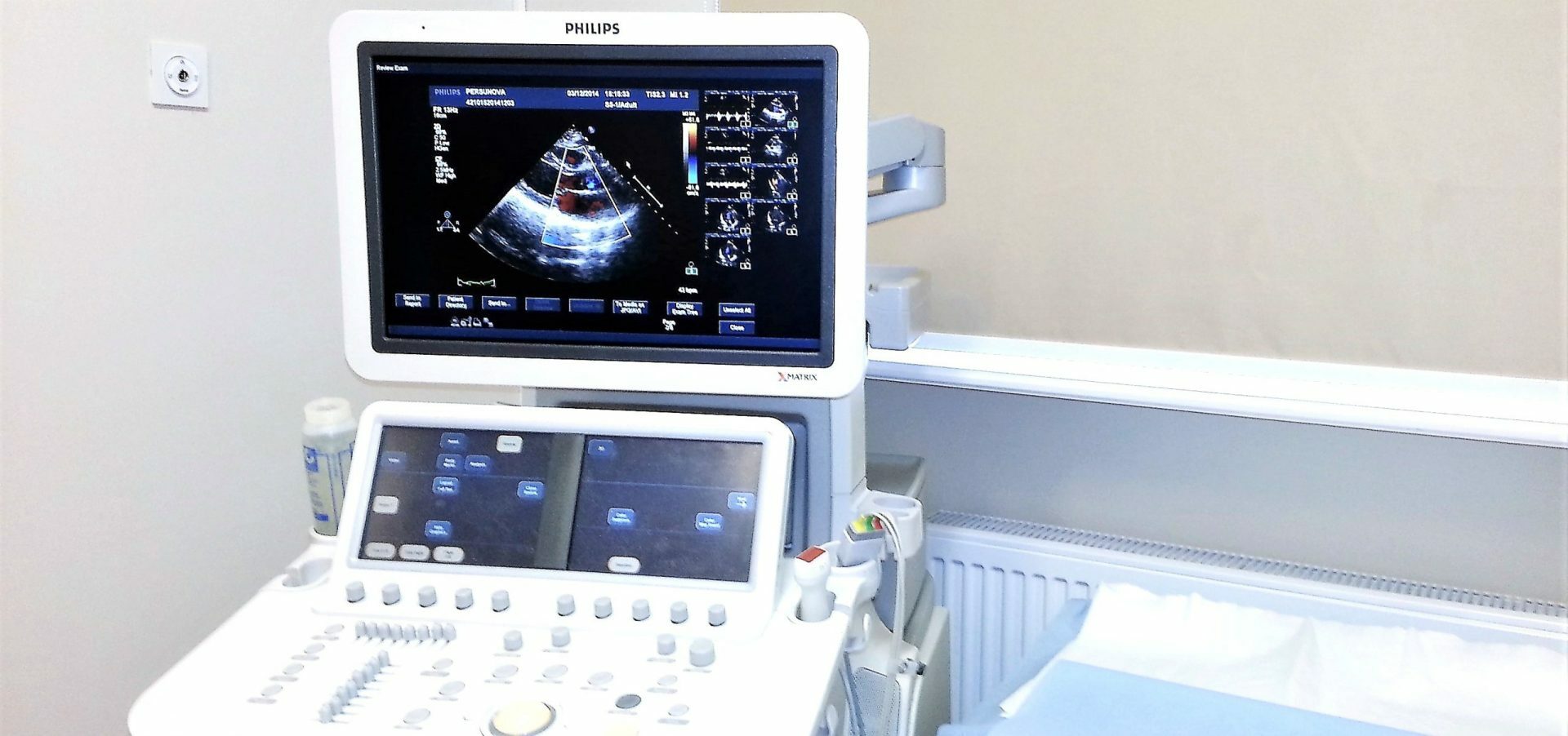
During the procedure, the patient lies down, and the doctor uses a probe to examine the heart, measure its size, evaluate function, and assess valve performance. The doctor also listens to the sounds of the heart valves and major blood vessels. A stethoscope is used to detect heart sounds and murmurs. The examination is painless, requires no prior preparation, and is completely safe. It can also be performed during pregnancy.
The procedure is conducted by a cardiologist who specializes in echocardiography. You can meet Riga 1st Hospital’s echocardiography specialists [here].
Other related services