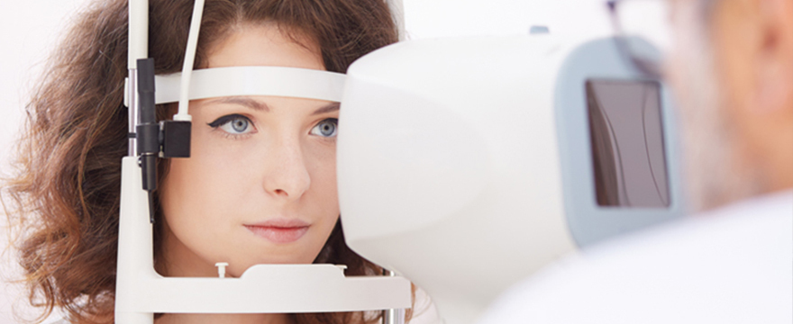

Ultrasound diagnostics (ultrasonography) is a safe, painless, highly informative, and quick visual diagnostic method used to assess the structure of various organs and soft tissues. High-frequency sound waves – ultrasound – are used to create the images.
Ultrasonography is increasingly chosen as an additional diagnostic method to clarify or monitor diseases. It is relatively precise, comfortable for the patient, and typically requires no special preparation.
When performing an abdominal ultrasound, it is possible to diagnose stones (gallstones and kidney stones), congenital or acquired organ anomalies, and even detect early signs of malignant processes in any organ.
Ultrasound can also determine:
- Causes of pain, menstrual irregularities, and bleeding; it can also evaluate male organs (testes and prostate) and female organs (ovaries and uterus), which is crucial in infertility cases.
- Fatty liver, chronic hepatitis, cirrhosis, and localized inflammatory foci such as abscesses, liver tumors, or metastases.
- Kidney cysts or solitary formations, kidney stones, congenital kidney pathologies, and whether there is congestion in the renal pelvis.
- Internal organ ruptures, bleeding in the organs or abdominal cavity, or free air in the abdomen, which may indicate stomach or intestinal perforation.
- Abdominal aortic aneurysms, where the aorta expands, often detected during the examination.
Ultrasound-guided biopsies can also be performed on various organs.
Ultrasound diagnostics at Riga 1st Hospital
At Riga 1st Hospital, experienced ultrasound specialists use the latest generation equipment to ensure precise and high-quality diagnostics.
Available ultrasound examinations include:
- Abdominal ultrasound – liver, gallbladder, pancreas, kidneys, spleen, lymph nodes
- Urological ultrasound – prostate (including transrectal), bladder, kidneys
- Thyroid and salivary gland ultrasound
- Testicular ultrasound
- Doppler ultrasound of the head and neck vessels
- Gynecological ultrasound (including for pregnant women and fetus)
- Echocardiography (heart ECHO)
- Doppler ultrasound of leg vessels
- Musculoskeletal ultrasound
- Breast ultrasound
- Transrectal 3D ultrasound
A doctor’s referral is required for state-funded ultrasound (USG) examinations. For paid services, a referral is recommended but not mandatory. The examination will be more targeted if conducted with a referral that includes information about the specific issue.
All ultrasound results are added to the e-platform (datamed), making it easy for other specialists to view the report, facilitating the transfer of medical documentation and reducing unnecessary repeat examinations.
Important: Insurance companies cover ultrasound examinations only if a doctor’s referral is provided.
Gynecological ultrasound at Riga 1st Hospital
Gynecological USG is a diagnostic method that uses ultrasound waves to visualize and assess a woman’s pelvic organs. It is a non-invasive and safe examination performed by a certified gynecologist or obstetrician in a clinic that meets regulatory requirements.
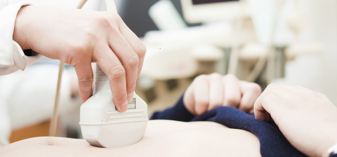
Abdominal ultrasound at Riga 1st Hospital
An abdominal ultrasound is a diagnostic procedure using sound waves to capture images of internal abdominal organs. It provides detailed information about organ structure, size, and function, helping identify possible pathological conditions or diseases.
Preparation for abdominal ultrasound:
- Avoid eating raw vegetables, fruits, or fruit juices for two days before the procedure.
- Arrive for the exam on an empty stomach (if the exam is after 3:00 PM, a light breakfast is allowed in the morning).
- Water and tea can be consumed without restrictions.
- All prescribed medications can be taken.
- If the ultrasound involves the urinary tract, bladder, or prostate, arrive with a full bladder, preferably not urinating for about three hours before the exam.
Take advantage of ultrasound diagnostics in a modern environment – trust Riga 1st Hospital!
Other related services