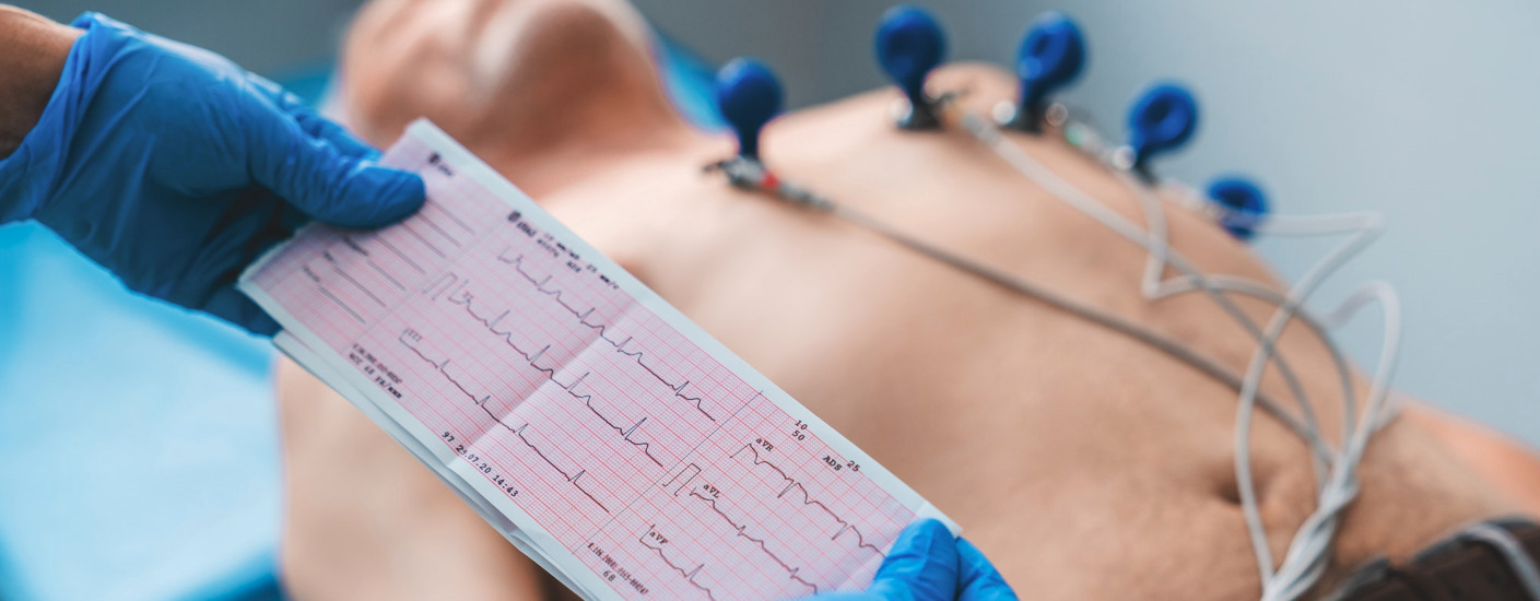
Electrocardiography: heart diagnostics and treatment
Electrocardiography (ECG) is a common method for diagnosing heart function. It is used to assess heart health, identify heart problems, and monitor the effectiveness of treatment.
ECG allows doctors to record the heart’s electrical signals and display them graphically as an electrocardiogram. This procedure evaluates heart function and can detect issues such as arrhythmias, heart ischemia (coronary disease), heart muscle damage, and other heart disorders.
An electrocardiogram is assessed alongside the patient’s medical history and other tests to determine the most appropriate and effective treatment.
When should an electrocardiogram be performed?
- During a routine check-up after an annual visit to a family doctor
- When a doctor suspects a new heart condition based on symptoms and examinations
- To evaluate an existing heart condition and the effects of medications
- Before a specialist consultation
- When heart diagnostics are needed for other medical conditions
- Before planned surgery
Heart examinations are essential for patients experiencing shortness of breath, weakness, swelling, palpitations, or chest pain. It’s best to perform these tests preventatively, before such symptoms arise.
The types of tests to be conducted are determined by the family doctor or cardiologist, who will assess the individual’s heart and cardiovascular risk (high cholesterol, diabetes, high blood pressure, smoking, etc.). Family medical history is also taken into account, such as whether close relatives have had heart or cardiovascular diseases.
How is electrocardiography performed?
The test requires no prior preparation and is painless. It can also be performed on children and pregnant women.
The test is done while the patient is lying down and electrodes are attached to the body. The electrodes are connected to the ECG device and attached to the patient’s skin to ensure an electrochemical connection between the heart and the device.
Medical staff record the electrocardiogram using an ECG machine. A standard ECG today has 12 leads, or electrodes, placed on different parts of the patient’s body to record electrical signals from the heart at various angles. Each of the 12 leads provides a different perspective of the heart’s activity, giving doctors detailed information about the heart’s condition.
What can an electrocardiogram reveal?
Electrocardiography is an effective method for evaluating heart rhythm and related parameters. It can detect:
- Acute coronary syndrome – a sudden manifestation of heart disease
- Various physiological conditions, such as anxiety, smoking, obesity, carbohydrate-rich meals before the test, pregnancy, etc.
- High blood pressure, heart chamber enlargement, and other conditions
Changes in the ECG can also be caused by non-heart-related conditions, such as kidney failure or thyroid diseases. If the patient has multiple conditions requiring specialist consultation and medication, all medications should be shown to the family doctor and specialist, as simultaneous use of different medications can affect ECG parameters and lead to serious heart rhythm disturbances.
Sometimes, rhythm disturbances are temporary and may not appear during the ECG recording. In such cases, the patient may be assigned long-term ECG recording, or Holter monitoring, which tracks heart activity for at least 24 hours under resting and active conditions. The recording period can be extended if necessary, or other monitoring methods may be used.
For diagnosing heart disease, assessing risk, prognosis, and treatment, a stress test with ECG, or an exercise stress test (ergometry), is used. It is conducted in a specially equipped room by a certified doctor and trained nurse.
Other related services


