
Modern and safe diagnostic method
Magnetic resonance imaging (MRI) is a modern visual diagnostic technique used to obtain images of various tissues through the use of a powerful magnetic field and radio waves. A radiologist interprets these images, identifying any tissue changes and making a diagnosis. This diagnostic method is a crucial part of patient healthcare, and Riga 1st Hospital offers this service with high-quality equipment and expert guidance. It is important to note that MRI is a painless and harmless procedure for the human body.
What examinations are performed?
MRI is used to examine various parts of the body and organs, including:
- Brain and spinal cord (central nervous system)
- Face, skull base, neck tissues, including orbits, salivary glands, and jaw joints
- Muscles, bones, and joints, including the spine
- Abdominal and pelvic organs
- Blood vessels
- Breasts
The primary goal of MRI is to provide an accurate and reliable diagnosis, which is essential for developing the patient’s treatment plan.
Fast, high-quality, and safe for patients
At Riga 1st Hospital, MRI examinations are performed by an exceptional team of professionals, led by Professor Ardis Platkājs, the Head of the Radiology Clinic.
“Radiology is a medical specialty that requires not only a deep knowledge of human anatomy and various disease manifestations but also the ability to use the latest information technologies and data processing systems,” says Professor Platkājs.
“At Riga 1st Hospital, we have gathered a team of outstanding professionals, which allows us to perform examinations quickly, efficiently, and safely for patients.”
“Many people, when coming for an MRI, as with other radiological examinations, are concerned about radiation exposure. However, the most important feature of MRI is the absence of ionizing radiation. MRI only produces radio pulses, which are commonly encountered in everyday life, such as when using mobile phones.”

Comprehensive diagnostic capabilities
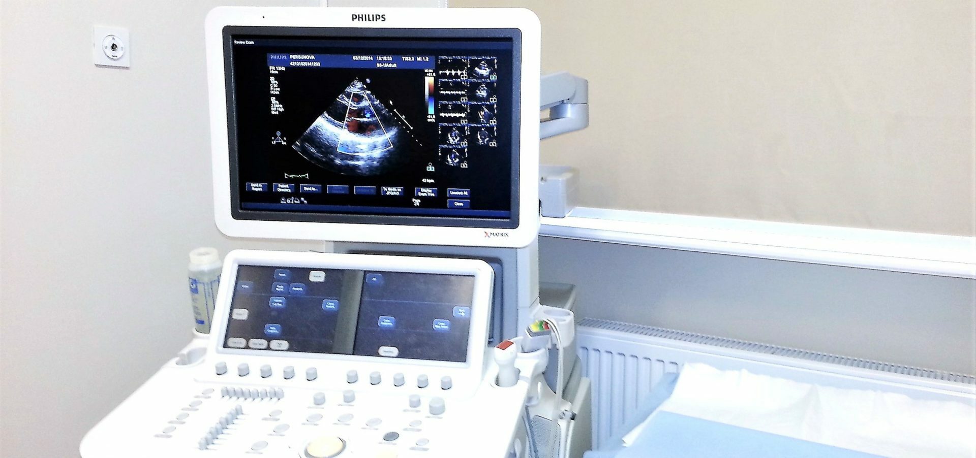
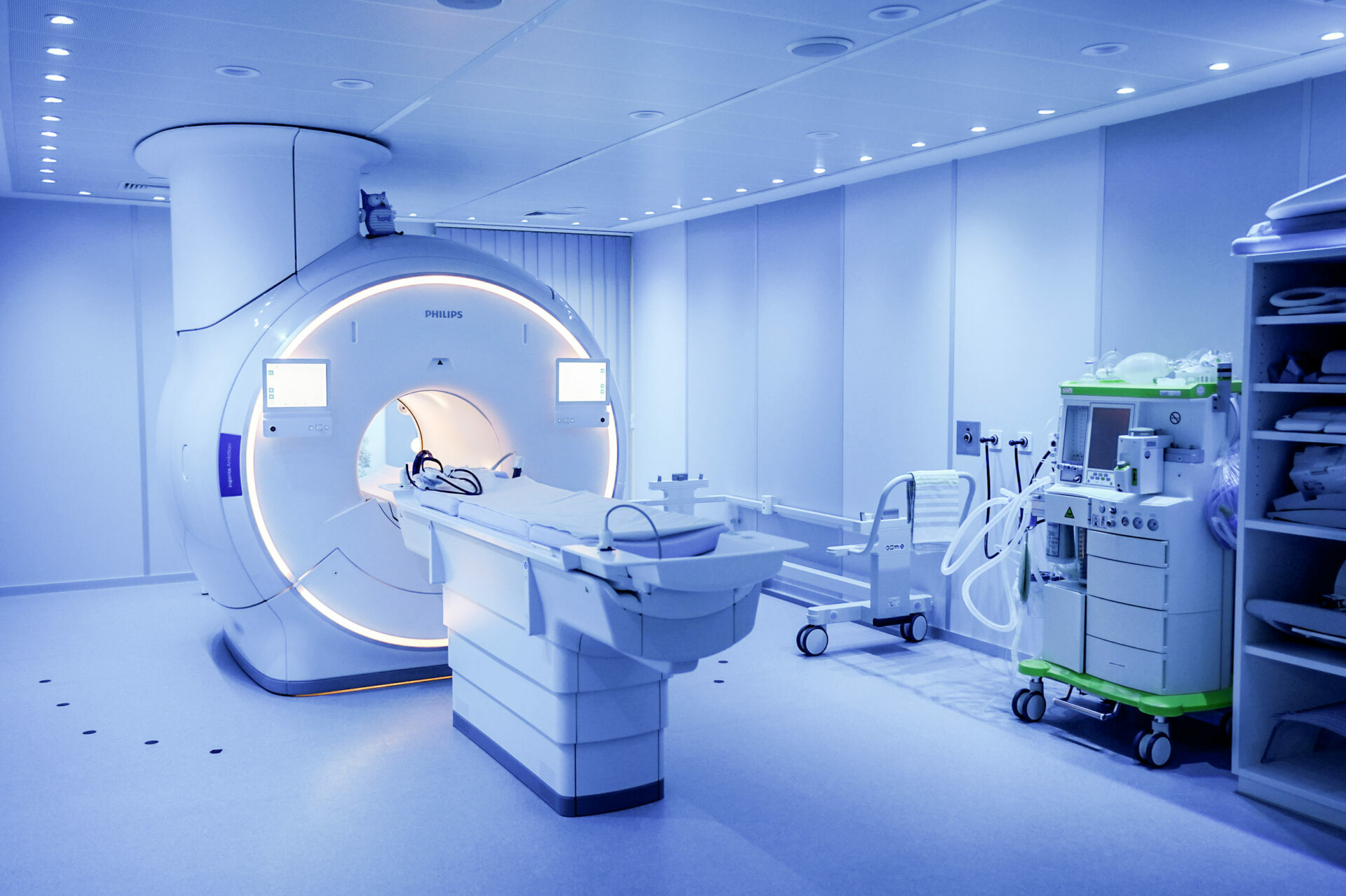
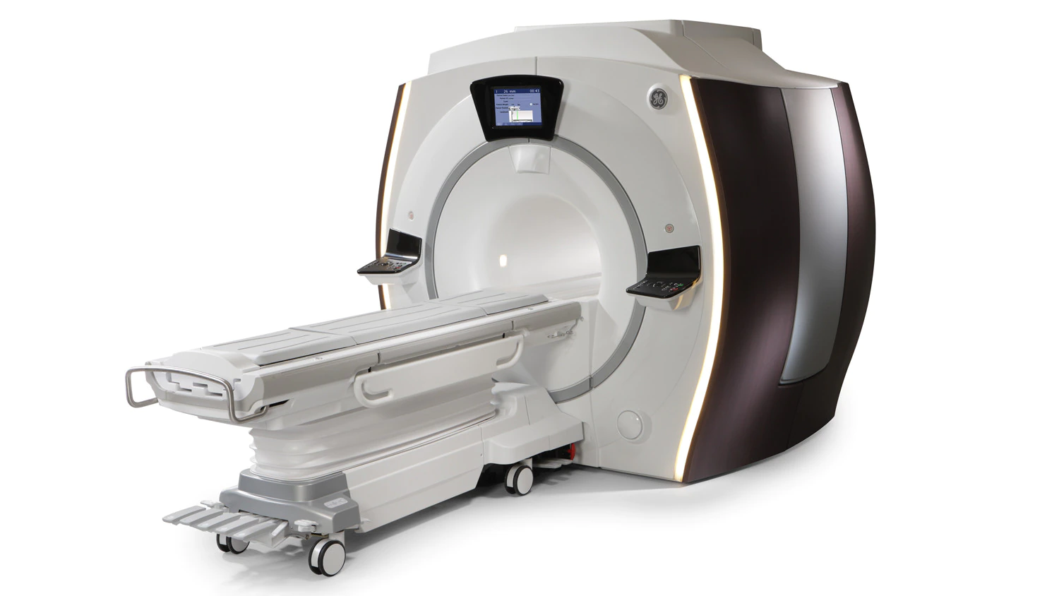
At Riga 1st Hospital, MRI is performed using two different machines, each with distinct technical features. This ensures comprehensive diagnostic options tailored to various patient needs.
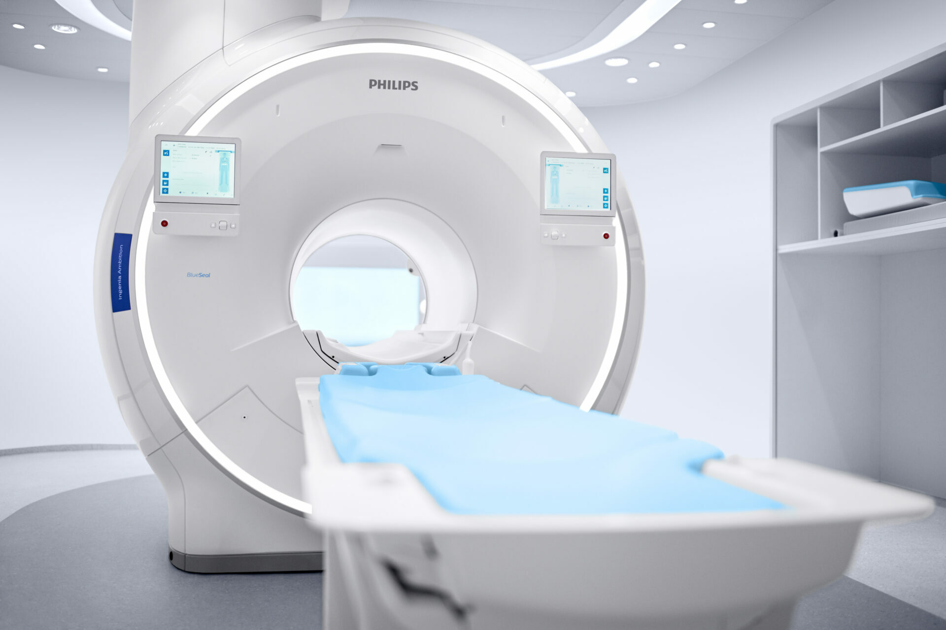
General Electric Healthcare (GE) MRI 3.0T
- Magnetic field strength: 3T (tesla)
- Highest resolution quality and examination speed
- The most advanced and informative examination programs
- Suitable for patients weighing up to 150 kg
Important information before the examination
To ensure a safe and effective MRI examination, it is crucial to inform the doctor who referred the patient about any of the following in the body:
- Pacemakers
- Neurostimulators
- Hearing implants
- Artificial heart valves
- Aneurysm clips
- Other metallic implants or objects
- Metal foreign bodies (bullets, shrapnel, metal clips, etc.)
- Pregnancy
Patients with metal implants
The decision regarding MRI for patients with metal implants is individualized and will be determined based on the specific circumstances.
MAGNETIC RESONANCE: FREQUENTLY ASKED QUESTIONS
- Pre-examination survey: Before the MRI scan, you will need to complete a survey where you must specify any metal objects in your body, such as post-traumatic loose metal fragments, post-surgical clips, screws, metal plates, endoprostheses, and others, as well as cardio-, neuro-, bone growth, or other stimulators. This is important because a strong magnetic field can affect metal objects and stimulators. The decision to accept a patient for MRI is made by a radiology assistant or radiographer.
- Claustrophobia: Since the patient must lie in a narrow tunnel (70 cm diameter) for 20-40 minutes, it is important to inform the radiology assistant or radiographer of any possible fear of enclosed spaces (claustrophobia).
- Allergies: If intravenous contrast agent administration is planned, inform the radiology assistant or radiographer of any allergic reactions to medications. Before the contrast agent is administered, it is recommended to check kidney function, such as glomerular filtration rate, urea, and creatinine levels in the blood.
- Dietary restrictions: For abdominal scans, avoid eating at least two hours before the examination. For pelvic organ and intestinal examinations, ensure the bowel is cleared of stool.
- Metal objects: Remove all metal objects, such as earrings, necklaces, piercings, etc., before the procedure. These items should be left in a special locker in the changing room. Wallets, phones, credit cards, and other valuables should also be left in the locker, which must be locked.
- Changing and protective covers: Patients should undress down to their underwear and put on a disposable gown, with protective shoe covers (bahilas) on their feet. Clothes should be left in the changing room, and the locker should be locked, with the key taken to the exam room.
- Earplugs: Earplugs will be offered to protect you from the noise generated by the MRI machine’s switches during the procedure.
- After the MRI examination, patients generally do not experience any changes in their well-being.
- If intravenous contrast was used during the examination, it is important to drink plenty of fluids to help flush the contrast agent from your body through urination.
- Magnetic resonance imaging (MRI) is well known for its safety and minimal risk of complications, so complications are rarely observed.
- When an MRI is performed with intravenous contrast, mild allergic reactions may occasionally occur, such as nausea, increased heart rate, or redness of the face or other body parts. However, these reactions are rare and generally not severe.
- The examination results will be available remotely, so the patient will not need to return to the hospital.
MAGNETIC RESONANCE IMAGING: WORKING HOURS
Examinations are available:
- Every weekday from 8:00 AM to 8:00 PM
- Saturdays from 8:00 AM to 2:00 PM
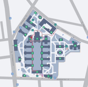
How to find the Department of Magnetic Resonance?
Other related services