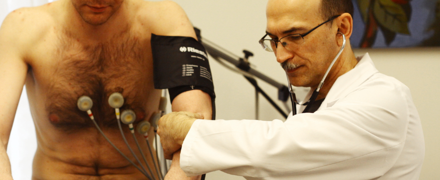


Dopplerography is a non-invasive method aimed at examining blood vessels using ultrasound technology. This examination provides comprehensive information about the speed and direction of blood flow in the vessels, allowing for the diagnosis of changes. Dopplerography can help detect vascular narrowing, dilation, as well as congenital conditions.
Dopplerography causes no complications and can be performed during pregnancy.
Dopplerography can be used to examine blood vessel conditions in the following cases:
- Various congenital developmental peculiarities, such as small-diameter vessels, vascular tortuosity, loops, or additional vascular branching
- Acquired vascular changes over a lifetime, such as arterial wall thickening, atherosclerotic plaques, or vascular narrowing or closures due to atherosclerosis
When is head and neck Dopplerography needed?
Head and neck vascular diagnostics are recommended for patients experiencing the following symptoms:
- Headaches, dizziness, and nausea
- Tinnitus and instability
- Headaches after physical exertion
- Blood pressure fluctuations
- High cholesterol levels in the blood
During head and neck Dopplerography, ultrasound equipment generates images of the blood vessels and records the direction and speed of blood flow within the vessel’s diameter. Dopplerography is recommended for the primary evaluation of the condition of the body’s arteries.
At the Diagnostic Radiology Clinic of Riga 1st Hospital, head and neck vascular duplex scanning with color-coded Dopplerography is available.
A diagnostic method that helps identify vascular problems at an early stage
Today, cardiovascular diseases, particularly strokes, are among the most common health threats. The main cause of strokes is atherosclerosis of the neck and brain blood vessels, which occurs when the vessels become clogged with cholesterol deposits. This is difficult to detect in the early stages, as the problem usually develops unnoticed. However, it can lead to serious consequences, such as paralysis, speech difficulties, intellectual impairments, and other issues.
To fully assess brain blood flow, Dopplerography is performed on both the neck and head vessels, as significant changes are often found in the neck vessels that branch out and supply blood to the brain.
Cervical brachiocephalic Dopplerography evaluates the anatomy of the neck arteries and identifies possible atherosclerotic changes in the arterial walls as well as changes in blood flow. Due to the easy access to the neck arteries, this is a very informative and important examination for all patients suspected of having atherosclerotic changes in the heart’s blood vessels.
What is atherosclerosis, and how does it affect health?
Atherosclerosis is a disease in which blood vessel walls thicken and lose elasticity. This occurs when fats accumulate inside the blood vessels, forming atherosclerotic plaques. These plaques narrow the blood vessels and disrupt blood flow, increasing the risk of stroke. Therefore, it is important to diagnose and treat it early under the supervision of a cardiologist, neurologist, family doctor, or vascular surgeon.
During Dopplerography, changes may be detected that require further, more detailed examinations using angiography methods (CTA, MRA, DSA). These examinations involve the use of contrast agents and either X-ray radiation or a magnetic field.
Important recommendations for improving health
To reduce the risk of stroke, it is essential to follow a healthy lifestyle and take care of your vascular health. Children should also learn to eat healthily and engage in physical activities from an early age. This will help prevent future vascular problems.
If you suspect vascular issues, consult a specialist and undergo a Dopplerography examination. This step will help detect and prevent problems early, reducing the risk of stroke.
We encourage patients planning to engage in physical activities to undergo Dopplerography to rule out risks that may arise from sudden head movements.
Other related services