
Computed tomography (CT): a visual diagnostic method
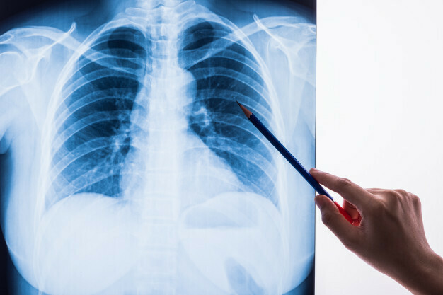
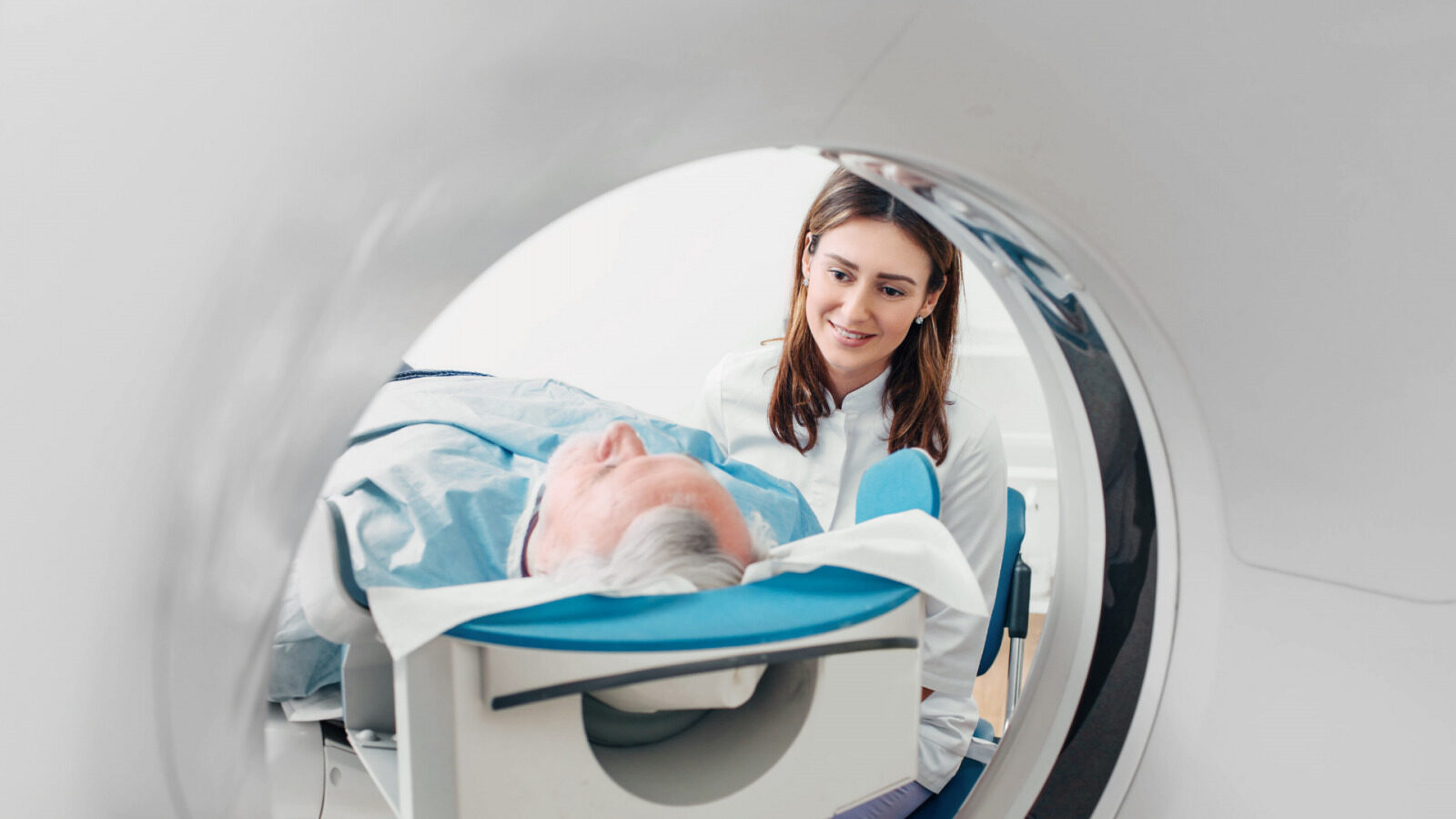
Computed tomography is one of the most important visual diagnostic methods in medicine, based on the ability of ionizing radiation to be absorbed by tissues. This diagnostic method provides high-quality cross-sectional images of the body, giving doctors the necessary information for studying and treating patients’ health.
High-quality body images and the latest technologies in healthcare
A computed tomography image is obtained through a process called reconstruction, which means digitally combining information from multiple projections at different angles in the examination area. During the procedure, an X-ray tube and detector rotate around the patient, creating highly detailed black-and-white images with a layer thickness of less than 1 mm. These images show internal organs and bones in two-dimensional form, with contrast depending on the attenuation of the rays during the examination. These differences allow radiologists to detect shadows that may indicate not only inflammation but also formations. The obtained CT images enable doctors to assess the anatomical features of organs, blood vessels, and other structures, their relationships, and the spread of pathological processes.
What examinations are performed?
Computed tomography is a versatile diagnostic method that can be used for any part of the body. Available at Riga 1st Hospital are:
- Brain examinations
- Angiography for evaluating the head’s blood vessels
- Sinus examinations
- Eye orbit examinations
- Temporal bone examinations
- Soft tissue examinations of the neck, including the larynx, thyroid, and others
- Angiography of the neck blood vessels
- Lung and mediastinum examinations
- Coronary and pulmonary artery angiography
- Liver, gallbladder, pancreas, spleen, adrenal gland, kidney, and lymph node examinations
- Abdominal and renal artery angiography
- Pelvic organ examinations
- Spine, cervical, thoracic, lumbar, sacral, and coccygeal bone examinations
- Full skeleton bone and joint examinations, including soft tissues
Computed tomography and bone examinations
CT is especially useful for bone examinations, as it accurately depicts the cortical bone (the hard part of the bone) and allows precise evaluation of foreign bodies, bone fragments, and splinters. The examination can be performed on patients with implanted medical devices such as pacemakers, magnetic vascular clips, or nerve stimulators.
Multislice computed tomography and organ disease diagnostics
Multislice CT is an essential examination for diagnosing liver, bile duct, and pancreatic diseases. The speed of this technology allows for quick examination at various time intervals and the evaluation of abdominal blood vessels and liver blood supply. The accuracy of the examination is enhanced by the use of a contrast agent injected into the patient. This examination is most commonly used to evaluate liver formations detected during previous examinations.
Multislice CT also provides high-quality, multi-planar 3D images of the pancreas, which play a crucial role in diagnosing pancreatic cancer and planning surgical treatment.
Use of contrast agents in computed tomography
Often, CT scans require the administration of a contrast agent. This substance, which contains iodine, helps improve the contrast of the images in the X-rays. Water-soluble contrast agents are typically used in vascular studies to evaluate blood supply and organ anatomy. If the patient has had allergic reactions to contrast agents or known kidney function disorders, it is important for the family doctor to provide this information.
The latest technologies reduce radiation risk during the examination
It is important to note that during a CT scan, the patient is exposed to ionizing radiation. The latest generation of CT equipment is designed to minimize radiation dose while maintaining diagnostically valuable images. These machines can reduce radiation by 40-80%, ensuring a safe and effective examination. However, the examination is not performed on pregnant patients.
The best 3D images of the heart and blood vessels
The latest of the two CT scanners at Riga 1st Hospital is the Discovery CT750 HD FREEdom by General Electric, offering the industry’s best spatial resolution for heart and vascular imaging. This device expands the possibilities for high-quality examinations of the head, spine, extremities, lungs, abdomen, pelvis, neck vessels, and coronary arteries.
Advanced programs provide new opportunities for examinations:
- For patients with metal prostheses (MARS – metal artifact reduction software)
- Kidney stone diagnostics
- Lung screening for early lung cancer diagnosis – for high-risk patients or smokers with low radiation exposure. This uses the LOW dose program with ASiR reconstruction (Advanced Statistical Iterative Reconstruction).
Highly qualified specialists with extensive experience
At Riga 1st Hospital, the team of experienced radiologists is led by Dr. Ard Platkajs, Ph.D., head of the Diagnostic Radiology Clinic.
Rasma Afremoviča is our senior radiographer. Her experience in diagnostics has been accumulated during her 33 years at Riga 1st Hospital. She communicates with patients daily, kindly and patiently explains the diagnostic process, administers contrast agents, and creates images of the examined organ or body part using the CT scanner.
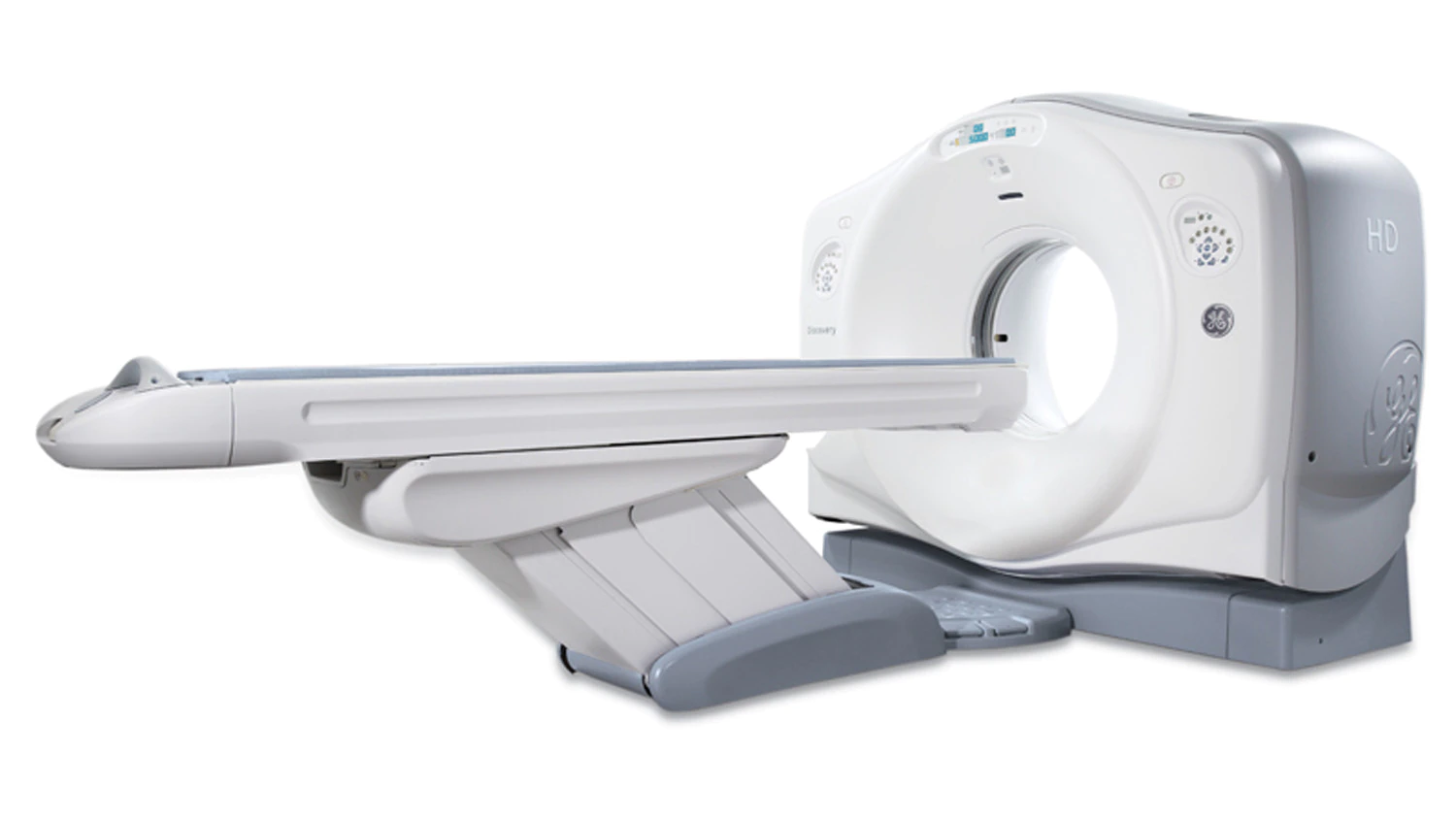
GE Revolution EVO Gen 3
- 64 detectors capable of generating 128 slices in one rotation.
- Enhanced imaging quality
- Faster scanning time
- Reduced radiation dose
- Improved patient accessibility
Maximum patient weight: 227 kg
Images from the device are automatically uploaded to datamed.lv.
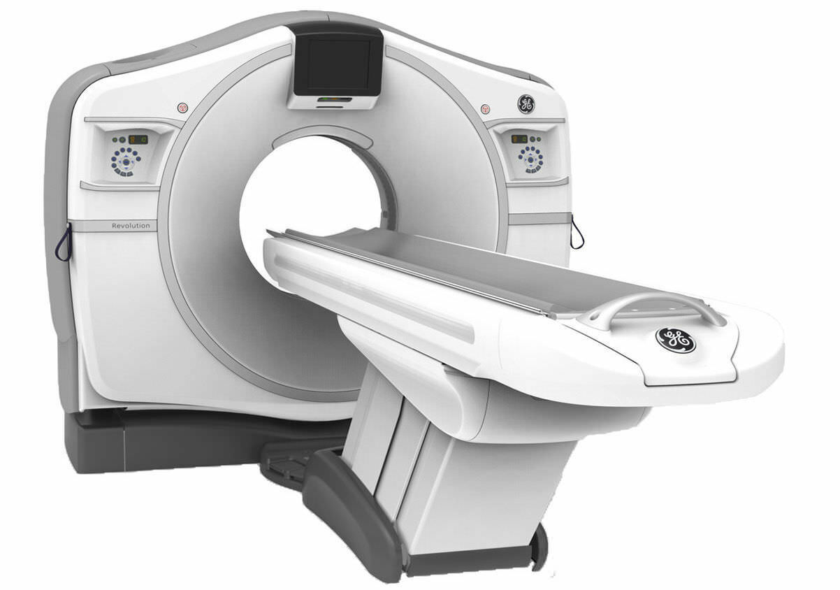
GE Discovery CT750 HD
- 64 detectors capable of generating 64 slices in one rotation.
- Fast scanning time
- Reduced radiation dose
- Improved patient accessibility
Maximum patient weight: 227 kg
Images from the device are automatically uploaded to datamed.lv.
Other related services